


Medical Equipment
Jeju National University Hospital shares stories of love and hope.
Da Vinci Surgical System
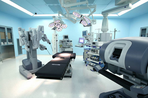
A surgical device recently touted as "Minimally Invasive Surgery". It minimizes scars and performs delicate surgeries more efficiently than the human. The robotic hands can rotate freely like human hands and surgeries can be performed safely without hand tremor. A surgery is perform in a non-abdominal operation state, leaving almost no scar. Such a surgery ensures less pain and fast recovery, and complications are less likely. The major surgical areas of the Da Vinci system are colorectal cancer, thyroid cancer, uterine myoma, ovarian cyst, uterine cancer, ovarian cancer, prostate cancer, bladder cancer, kidney cancer, laryngeal cancer, and oral cancer.
Varian/Rapid ARC
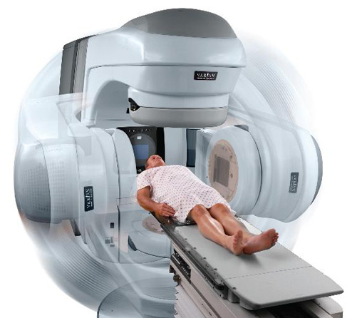
RapidArc is a piece of radiation therapy equipment that enables the latest VMAT (volumetric arc therapy). VMAT is similar to that of the existing Tomotherapy in that it irritates the entire tumor while rotating 360 degrees. However, contrary to that Tomotherapy uses a treatment method of dividing tumors into multiple monolayer, VMA treats the entire tumors at the same time, reducing overall treatment time. Unlike the conventional IMRT (Intensity-modulated Radiation Therapy) that radiates in the fixed angle, VMA reduces the amount of radiation that reaches the normal tissues around tumor while transferring radiation to tumors in the precise shape. With this, we can reduce side effects of radiation therapy while expecting to improve the therapeutic effect.
PET-CT - Siemens/Biograph True Point 40
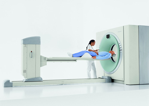
PET-CT is a system combined with PET that examines the abnormal metabolism of the body and CT that examines structural abnormalities. With excellent performance of prevention and early detection of cancer, the system examines precisely an incidence of cancer occurrence of the body through a single shot and detects invisible cancer cells. PET-CT is achieved by the fusion of positron tomopgraphy and 40-slice multi-direction computed tomography results thus is effective for brain and heart-related diseases, especially early diagnosis and treatment progress of cancer.
Cyclotron – Samyoung Unitech/KIRAMS / KIRAMS 13
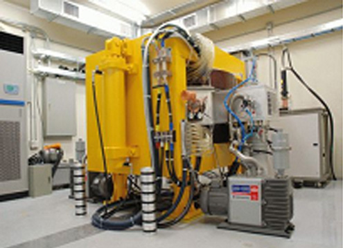
Cyclotron is a particle accelerator that accelerates particles and composite particle ion in the uniform magnetic field, and a system that generates positron-emitting radioisotopes used in Pet, the essential equipment for cancer diagnosis.
Multi-directional computed tomography - (MDCT)(Siemens / Somatom Definition)
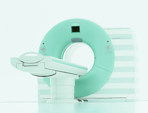
We are currently operating two units of MDCT, and a dual source MDCT (Siemens / Somatom Definition), recently introduced, is equipped with two detectors of which measures X rays that penetrated a patient and tubes in which generate X rays. The new device is twice more faster than the existing MDCT and ensures clearer images. Hence, the system will better help achieving imaging diagnosis of heart and cerebrovascular disease and emergency trauma patients that require rapid CT scans.
Magnetic Resonance Imaging Camcorder - (MRI)(Phillps / Achieva MR 3.0T)
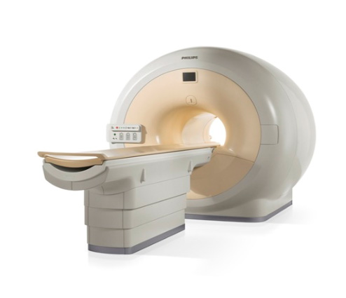
The hospital uses two units of MRI, where the camcorder is 1.5T of Siemens and 3.0T of Phillips. In particular, Phillips' new Achieva MR has reduced examination time and the burden of patients with emergency or narrow phobia, including patients being unable to self-control. It also minimized the amount of contrast media administered to patients and reduced the risk of side effects. With its high clarity, we were able to perform precise diagnoses of all the microstructure of the body and easily diagnose cerebral diseases such as epilepsy and dementia using special imaging techniques. In addition, we provide excellent information for measuring metabolite changes, early diagnoses, and treatment of tumor and metabolic diseases.
Ultrasound System
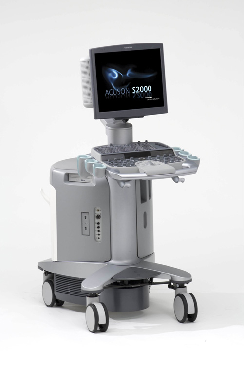
Radiology of the JNUH is equipped with four ultrasound scanners (Model Name: 1 unit of Siemens' Acuson S2000, 1 unit of Philips' HDI 5000, 1 unit of HDI sono CT, and 1 unit of ALOKA-prosound) for precise ultrasound diagnoses and our medical expertise are providing the best ultrasonic images. Professors of Radiology are conducting examination according to their detailed study fields, such as upper abdomen (liver, gallbladder, biliary tract, kidney, spleen, etc.), breast, thyroid, neck, musculoskeletal, and vascular color Doppler ultrasound.
Digital Vascular Camera - (Siemens - Artis zee / Philips- Allura xper FD2020)
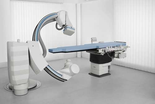
The JNUH is equipped with three digital vascular cameras. The latest cardiovascular imaging equipment인 "Artis zee" the latest cardiovascular imaging equipment of Siemens allow us to achieve very sharp 2Dl and 3D images while minimizing the patients' radiation exposure; hence, it allows heart physicians to determine acute myocardial infarction and angina pectoris, and accurate cardiovascular conditions of patients for rapid, accurate, and effective percutaneous coronary treatment. The latest angiographic apparatus "Allura xper FD2020" of Philips provides high-resolution images for the diagnosis of vascular disease, cerebrovascular and vascular intervention procedure of all parts and ensures effective and accurate diagnosis and treatment. Moreover, the design put importance on patients' safety by using contract media and minimum X rays, and supports accurate and effective treatment via 3D image simulation. The XperCT function that implements images similar with CT scanning ensures rapid diagnosis and treatment.
Digital Mammography

The device is the lasts model (Model Name: Mammomat Inspiration) of Siemens, which offers high-resolution images and dramatic reduction of the amount of X rays compared to other mammography systems. The system induces appropriate pressure and reduces homeopathic patients during scanning; in addition, the patient-friendly design and functions ensure comfortable scanning experience to patients.
Automatic chemistry analyzer - (Toshiba / TBA-200FR)
A cutting-edge system that produces accurate examination results of a variety of examination items (Max. 100 items /or items chosen by patient) within a shortest time by using a small amount of blood.
Digital Radiography System & Fluoroscopy System

Our Digital Radiography Systems consists of three latest model of Siemens (model name: 1 Axiom Aristos Vx 1, 2 Axiom Aristos Mx) and two units of fuoroscopy machine (model name: Axiom Iconos R200). The systems provide high-resolution images and they offer a digital batch processing from the registration of patients, examination to image transmission. This has decrease patients' waiting time, thereby offering comfortable examination experience.
Extracorporeal Shock Wave Crusher - (EDAP / TMS)

Contrary to the existing ureteral stones and kidney stones surgeries, the device crushes stones in the kidney, ureter and bladder and produces them in fine particles using high-energy shock waves and ensure they are excreted in the urine. The safest and most effective treatment does not require admission nor surgery or anesthesia..
Respirator - (Drager medical / Evita 4)

The device prevents infection by tube as it allows artificial respiration using masks without tube insertion, which is the old method of artificial breathing by inserting tubes into the airway. The operating state of the artificial respirator is shown on the screen ergonomically so as to check the respiration state of a patient and to provide the optimum oxygen level in order to effectively control the use of a respirator.
Digital Operating Room System - (Striker)
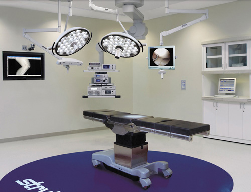
An operation room equipped with world-class competitiveness. With the system, we can transmit real-time surgical images between international conference rooms or the main conference of the hospital. The system ensures remote operation with the help of real-time medical advice during an operation participated by medical specialties around the world, professionals of local hospitals, and operation surgeon in the operation room.
StealthStation® S7™ and Fusion™ ENT Navigation System
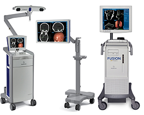
Electromagnetic intra-operative imaging systems of StealthStation® S7™ and Fusion™ ENT Navigation System are innovative equipments, enable surgeons to see the exact location and anatomical structure during surgery. These navigation system provides updated information with real-time images and offers the best patient care through brain tumor surgery, endoscopic skull base surgery, and endoscopic functional sinus surgery with accuracy and convenience to use.